What Best Describes the Structure of the Kidneys
-Each kidney is retroperitoneal and anchored by adventitia. This enzyme helps manage the expansion of arteries and the volumes of blood plasma lymph and interstitial fluid.

Renalnephrons Diagram Of Kidney And Nephron Kidney Anatomy Renal Physiology Anatomy And Physiology
Muscles in your back and your ribcage protect your kidneys from both the front and the back sides of your body.

. The kidneys are bilateral organs placed retroperitoneally in the upper left and right abdominal quadrants and are part of the urinary system. A thin connective tissue called the renal capsule surrounds each kidney. What best describes the main function of the kidneys.
They are the microscopic structure composed of a renal corpuscle and a renal tubule. Of the kidneys is just below the rib cage with one on each side of the spine. Describe the external structure of the kidney including its location support structures and covering.
A The glomerulus produces erythropoietin. Which of the following best describes the outcome if the kidneys stopped functioning. Each kidney is made up of over one million nephrons that dot the renal cortex giving it a granular appearance when sectioned sagittally.
Blood would fill with waste and the human body would not be able to maintain homeostasis. D it is superior to the uterus. The main function of the kidneys is to filter the blood to remove uric acid and other unwanted substances.
There are two types of nephrons cortical nephrons 85 percent which are deep in the renal cortex and juxtamedullary nephrons 15 percent which lie in the renal cortex close to the renal medulla. Kidney Structure The bean-shaped kidneys have an outer convex side and an inner concave side called the renal hilus where the renal artery vein and ureter are found. What structures are labelled I II and III in the diagram of the kidney.
A collection of tubelike structures that leads away from the glomerulus. Which statements correctly describe the positioning and anatomic structure of the kidneys in the abdominal cavity. The right kidney is placed slightly lower- than the left due to the presence of liver which occupies much space on the right side.
Identify the major internal divisions and structures of the kidney. This controls erythropoiesis which is the production of red blood cells. D The renal arteries arise from the renal cortex.
The liver also produces erythropoietin but the kidneys are. B The renal pelvises drain urine into the ureters. The left kidney sits a bit higher in the body because of the size of the liver which is also on the right side.
Their shape resembles a bean where we can describe the superior and inferior poles as well as the major convexity pointed laterally and the minor concavity pointed medially. Which of the following best describes the structure of the kidneys. A mass of capillaries that makes up the glomerulus.
A nephron is the basic structural and functional unit of the kidney. The kidneys would fill with urine d. Each kidney is about four or five inches long shaped somewhat like a bean.
The right kidney is generally slightly lower than the. Compare and contrast the cortical and juxtamedullary nephrons. B it is posterior to the kidneys.
This capsule maintains the kidneys shape and protects the inner tissues. The renal parenchyma can be divided into two main areas the outer cortex and inner medullaThe cortex extends into the medulla dividing it into triangular shapes these are known as renal pyramids. The apex of a renal pyramid is called a renal papillaEach renal papilla is associated with a structure known.
One on each side of the vertebral column at the level of the 12th thoracic to 3rd lumbar vertebrae. Two tubes that drain urea produced in the liver and transport it to the kidneys where it is filtered Two tubes that drain urine produced in the kidneys and transport it to the bladder where it is collected Two organs that collect urine produced in the kidneys until ready to be. The functional units of kidney.
The kidneys are two in number which are situated one on each side of the verteral column and in-front of the last ribs. Outside peritoneal cavity in the back of the upper abdomen. Which of the following statements best describes the structure and function of the ureters.
A renal corpuscle and a rena. Lymph is a fluid that. The human kidney is a reddish-brown bean-shaped structure situated between the last thoracic and third lumbar vertebra close to the dorsal inner wall of the abdominal cavity.
They are about 1114 cm in length 6 cm wide and 4 cm thick and are directly covered by a fibrous capsule composed of dense irregular connective tissue that helps to hold their shape and protect them. 2 what color does urine exhibit with elevated and takes a B in C vitamins. 1 Which is true of the uterus.
The nephron is the kidneys microscopic structural and functional unit. Paired multilobar up to 18 each lobule contains nephrons. C it is inferior to the bladder.
There are about millions of nephrons in each human kidney. Identify the major blood vessels associated with the kidney and trace the path of blood through the kidney. Internally the kidneys have an intricate and unique structure.
Blood would increase its carbon dioxide concentration b. Each kidney weighs about 125175 g in males and 115155 g in females. The kidney is covered by fibrous connective tissue the renal capsule which protects the kidney Internally It consists of the outer dark cortex and an inner light medulla both containing nephron.
A it is superior to the bladder. Branches of the renal artery that supplies blood to the renal cortex. The frequency in which a human excretes urine would increase.
-The right kidney lies slightly lower than the left kidney on each side of the spinal column. The word nephron is derived from the Greek word nephros meaning kidney. -Each kidney has an outer cortex and inner medulla.
They lie on the posterior abdominal wall. Tubelike structures that converge to form the ureter. C The cortical nephrons concentrate urine.
Terms in this set 39 structure.

The Kidney Kidney Anatomy Anatomy And Physiology Human Kidney

Kidney Structure Biology For Majors Ii

Kidney Structure Biology For Majors Ii
Kidney Anatomy Nephron Filtration Diagram Photo Kidney Anatomy Basic Anatomy And Physiology Anatomy

Anatomy Study Cushings Syndrome Home Remedies For Uti Polycystic Kidney Disease

Effective Tips For Maintaining Good Kidney Health Kidney Anatomy Human Body Organs Human Anatomy And Physiology

Kidney Anatomy Cross Section Image Kidney Anatomy Anatomy Images Arteries Anatomy

Kidney Structure Biology For Majors Ii

25 1 Internal And External Anatomy Of The Kidney Anatomy Physiology

Gross Anatomy Of The Kidney Anatomy And Physiology Openstax Renal Physiology Kidney Anatomy Renal

Realistic Human Internal Organs Infographics White Background With Lungs Heard Liver Kidneys Brain Eyes Spleen Intestines D Human Body Organs Infographic Human

Major Calyx Minor Calyx Renal Capsule Anatomy In The Kidney Www Anatomynote Com Renal Kidney Anatomy Kidney Cyst
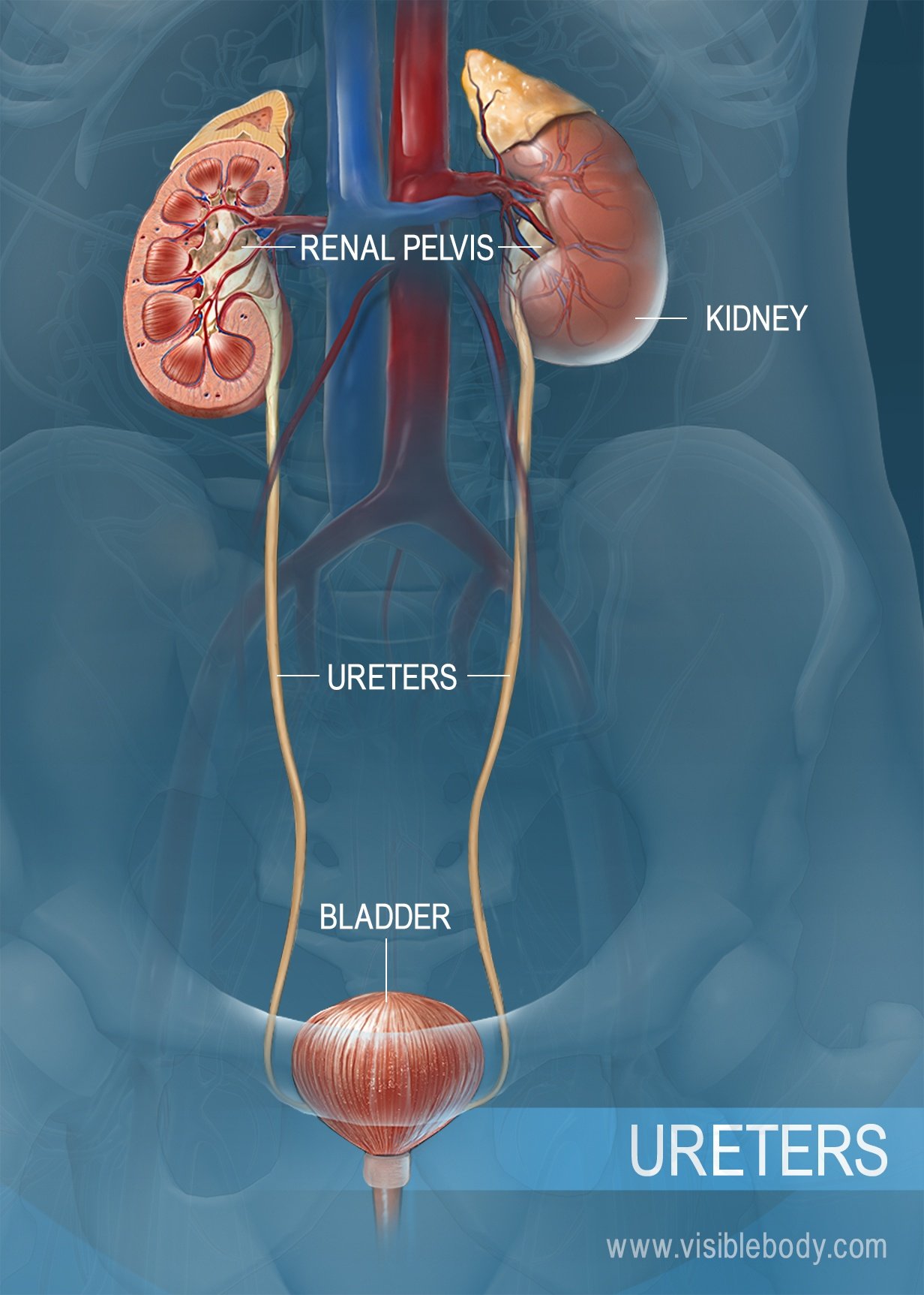
Urinary System Structures

Pin On Sistema Excretor

Pin On Nursing

Pin On A P

The Urinary Tract Chart 20x26 Medicinskie Uchilisha Anatomiya Cheloveka Plakat

Pin On Veterinary Online

The Kidney Chart 22x28 Kidney Anatomy Kidney Human Body Muscles


Comments
Post a Comment